
Text: Natalie Bakir, Matthieu Beaugrand, Salome Kurth
As children progress through their developmental journey, encompassing various facets such as cognitive, emotional, and physical growth, a remarkable phenomenon comes to light. In their deep sleep electroencephalogram (EEG) a progressive shift occurs in the localization of slow-wave activity (SWA) which moves from posterior to anterior regions. Importantly, these maps of SWA are connected to essential neurobehavioral functions in childhood and adolescence, for example motor skills. Yet the connection between sleep maps during the early period of life – infancy – and subsequent behavioral outcomes remains uncertain.
The investigation of how behavioral development and infant sleep EEG maps are linked is a subject of significant interest, which could provide insights into the intricate early developmental processes.
Our latest investigation tested whether healthy infants’ brain activity during the first hours of nocturnal sleep is linked to behavioral scores and developmental outcomes.
Specifically, we wanted to test whether complex sleep maps (based on over 100 electrodes) can be used in a simplified way to predict behavior. We reduced the number of electrodes to the ones over brain regions that undergo the most substantial maturational dynamics – these are located over frontal, central and posterior regions of the scalp.
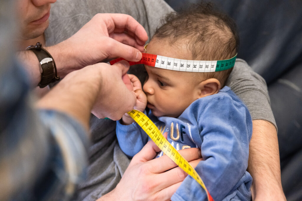
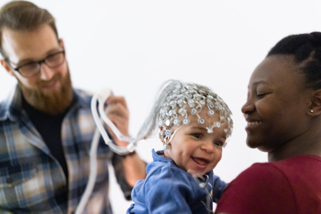
In a longitudinal study by Matthieu Beaugrand et al. (1) a total of 35 healthy 6-month-old infants were measured with high-resolution EEG (see photo) in a sleep assessment at the families homes. Parents completed surveys regarding the infants’ behavior at the ages of 3, 6, 12, and 24 months.
The results show that simplified maps of EEG power in healthy infants age 6 months do NOT directly correlate with behavioral development at 3, 6, 12, or 24 months, contrary to previous findings in older children (2)(3) or clinical cohorts (4). In contrast, in a separate analysis published recently, we identified spindle density as a potential early marker for infant behavioral developmental outcomes (5).
There is thus a need for further examination of the complex relationship between developmental processes and the neurophysiology of sleep specifically at early age. Only longitudinal assessments starting as early as possible will enhance our understanding of how brain wiring matures.
Sources
- Beaugrand, M., Jaramillo, V., Markovic, A., Huber, R., Kohler, M., Schoch, S., Kurth, S. (2023) Lack of association between behavioral development and simplified topographical markers of the sleep EEG in infancy, Neurobiology of Sleep and Circadian Rhythms
- Kurth, S., Ringli, M., Geiger, A., LeBourgeois, M., Jenni, O. G., & Huber, R. (2010). Mapping of Cortical Activity in the First Two Decades of Life: A High-Density Sleep Electroencephalogram Study. Journal of Neuroscience, 30(40), 13211–13219.
- LeBourgeois, M. K., Dean, D. C., Deoni, S. C. L., Kohler, M., & Kurth, S. (2019). A simple sleep EEG marker in childhood predicts brain myelin 3.5 years later. NeuroImage, 199, 342–350.
- Guyer, C., Werner, H., Wehrle, F., Bölsterli, B. K., Hagmann, C., Jenni, O. G., & Huber, R. (2019). Brain maturation in the first 3 months of life, measured by electroencephalogram: A comparison between preterm and term-born infants. Clinical Neurophysiol
- Jaramillo, V., Schoch, S., Markovic, A., Kohler, M., Huber, R., Lustenberger, C., & Kurth, S. (2023). An infant sleep electroencephalographic marker of thalamocortical connectivity predicts behavioral outcome in late infancy. NeuroImage, 269, 119924.
Thanks
Photo: Minnie Zhou, Unsplash
Image Stemutz
ChatGPT for texting a first draft